







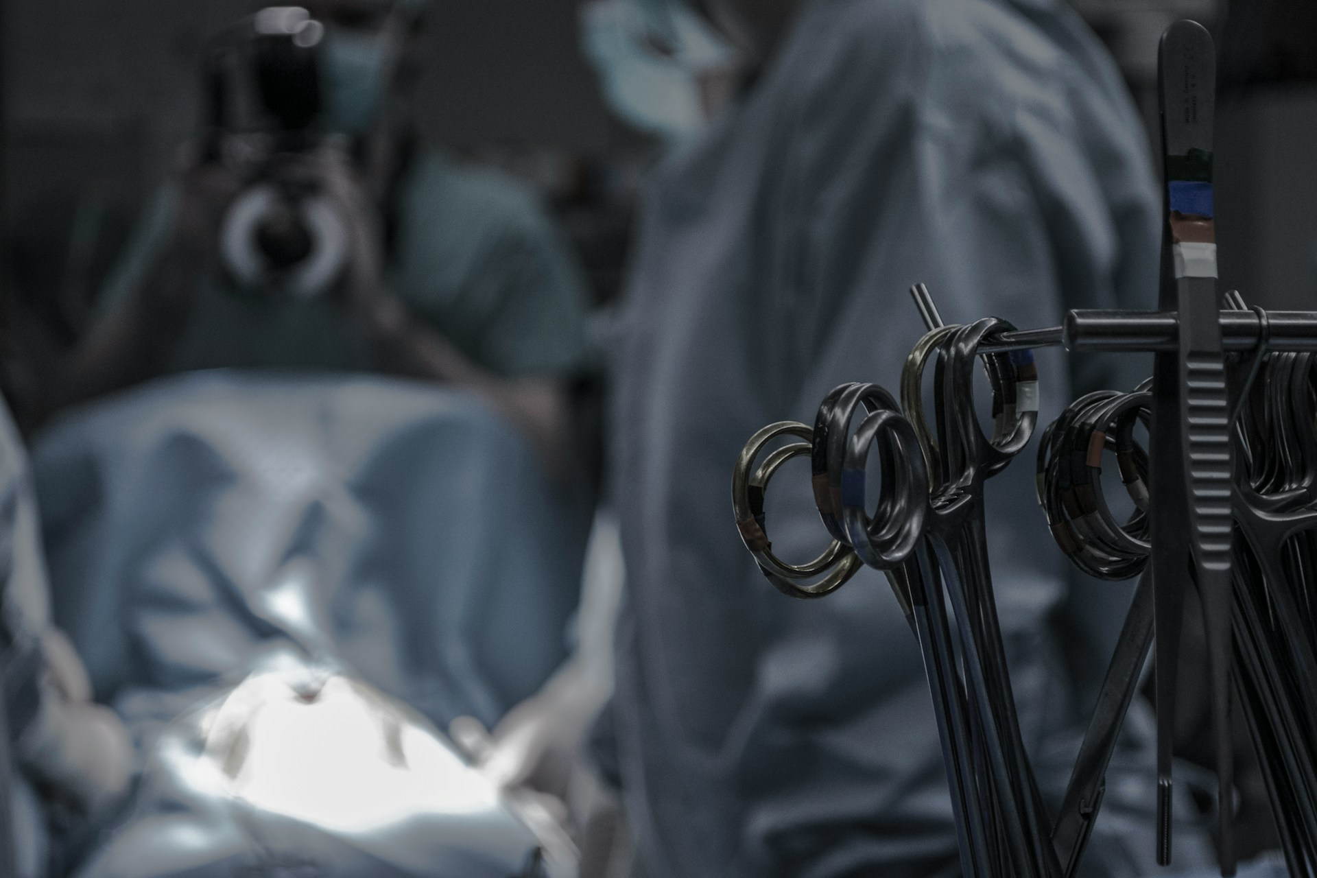
Persistent back pain radiating down your leg or chronic neck discomfort that makes turning your head agonizing often points to a single culprit: disc herniation. For years, the prospect of spine surgery brought forward anxieties about large incisions, significant muscle disruption, and prolonged bed rest. Advances in surgical technology have dramatically shifted this narrative. Today, minimally invasive spine procedures enable surgeons to treat herniations with millimeter-level precision while leaving surrounding tissues largely undisturbed.
Understanding Disc Herniation: When Conservative Care Isn’t Enough
Intervertebral discs act as shock absorbers between your vertebrae, composed of a tough outer ring (annulus fibrosus) and a soft, gel-like interior (nucleus pulposus). When wear, sudden trauma, or chronic strain causes this outer ring to tear, the inner material can bulge outward, compressing sensitive nerve roots or the spinal cord itself.
Initial care typically involves conservative management: rest, physical therapy, targeted anti-inflammatory medications, and epidural injections. However, if progressive neurological deficits appear—such as persistent numbness, significant motor weakness in the leg or foot, or intractable pain resistant to medication—surgical decompression becomes essential to prevent permanent nerve injury.
Microendoscopic Discectomy: The Hybrid Microscopic & Endoscopic Approach
Microendoscopic discectomy serves as a powerful bridge between traditional microsurgery and modern endoscopy. In this technique, the surgeon localizes the exact spinal segment using intraoperative fluoroscopy (real-time X-ray guidance) and establishes access using progressive muscle-dilating tubes rather than cutting through structural back muscles.
-
Incision Profile: Typically requires a small opening of 2 to 4 cm.
-
Visualization: Combines the deep, multi-angle illumination of an endoscope with high-magnification surgical operating microscopes.
-
Clinical Utility: This dual setup gives the neurosurgeon exceptional depth perception, making it particularly advantageous for complex disc anatomies, revisions, or where localized bone removal is required alongside disc decompression.
Uniportal Spine Endoscopy: Keyhole Precision Through Sub-Centimeter Incisions
Uniportal endoscopy represents ultra-minimally invasive spine surgery. Instead of relying on larger surgical corridors, this technique routes all visualization channels and micro-instruments through a single, slender tubular sheath.
-
Incision Profile: Consistently measures less than 1 cm (frequently around 0.5 to 1 cm).
-
Mechanism: The surgeon works under continuous irrigation with saline, which clears away micro-bleeding, keeps the surgical field pristine, and gently separates delicate nerve tissues without traction.
-
Patient Advantage: Because muscular detachment is virtually zero, postoperative wound pain is negligible. Many patients are able to stand and walk within hours of the procedure.
Unilateral Biportal Endoscopy (UBE): Maximum Visualization with Minimal Tissue Disruption
Unilateral Biportal Endoscopy balances the visual reach of full endoscopy with the surgical dexterity of open surgery. Rather than combining the camera and instruments into a single port, UBE separates them into two independent portals on the ipsilateral (same) side of the spine.
-
Incision Profile: Two tiny entry points, each measuring roughly 1 to 2 cm, placed just centimeters apart.
-
Mechanism: One portal houses a high-definition endoscope providing panoramic visualization, while the second portal serves as dedicated access for surgical tools.
-
Key Benefits: Because the working instrument moves independently of the lens, the surgeon gains complete freedom of movement. This makes UBE exceptionally versatile for challenging angles, extensive canal stenoses, and bilateral decompression performed from a unilateral entry.
Beyond the Lower Back: Treating Cervical and Thoracic Disc Disorders
While lumbar (lower back) disc herniations are the most frequent cause of sciatica, identical pathology often affects other regions of the spine:
-
Cervical Spine (Neck): Herniations here trigger radiculopathy—sharp shooting pain, tingling, and weakness radiating down the shoulder, arm, and fingers. Endoscopic cervical approaches allow decompression without requiring major anterior neck dissection or spinal fusion in select patients.
-
Dorsal/Thoracic Spine (Mid-Back): Mid-back disc herniations are less common but technically demanding due to the proximity of the rib cage and the vulnerability of the thoracic spinal cord. Minimally invasive endoscopic approaches provide direct lateral access, decompressing the cord without the morbidity of traditional open chest surgery.
Recovery and Rehabilitation: Getting Back to Daily Life Faster
The advantages of smaller incisions extend far beyond cosmetic appearance. Minimal muscle dissection translates to:
-
Reduced Blood Loss: Minimal to no blood transfusion requirement.
-
Shorter Hospitalization: Most endoscopic patients return home within 24 to 48 hours, with select cases performed as outpatient day surgeries.
-
Faster Functional Recovery: With natural spinal architecture and ligaments preserved, structured physical rehabilitation can begin much earlier, enabling a safe and rapid return to desk work, light exercise, and everyday life.